Progression of Artificial Intelligence in Prenatal Ultrasound:
Advancing AI Applications in Fetal Heart Assessment
Martin Chavez, MD1, FACOG, FAIUM and Julia Kim, MD1
1Division of Maternal-Fetal Medicine, Department of Obstetrics and Gynecology, New York University Langone Hospital Long Island, New York University Grossman Long Island School of Medicine, Mineola, NY.
Introduction
The concept of artificial intelligence (AI) in the last few years has taken off in society at speeds not seen most likely since the industrial revolution. The advent of artificial intelligence (AI) is poised to have a transformative impact on society, comparable to the sweeping changes brought about by the Industrial Revolution over 200 years ago. However, while the Industrial Revolution primarily affected how tasks were performed mechanically, AI’s influence extends far beyond, fundamentally altering our cognitive capabilities and productivity1. AI has the potential to dramatically compress the time required for human achievements. Tasks that once took years or even decades could potentially be accomplished in a fraction of the time. This acceleration of progress is not limited to physical labor but extends to intellectual pursuits, creative endeavors, and complex problem-solving. Imagine a scenario where scientific breakthroughs that might have taken a century of cumulative human effort could be realized within a single generation. AI’s ability to process vast amounts of data, recognize patterns, and generate novel solutions at unprecedented speeds could catalyze advancements across various fields, from medicine and environmental science to space exploration and beyond.
As we embrace the possibilities of AI, it’s crucial to steer its development and application towards enhancing human potential rather than replacing it, ultimately aiming for a symbiotic relationship between human creativity and artificial intelligence. As maternal-fetal medicine specialists, we are captivated by the potential symbiosis between artificial intelligence and our field. Our daily practice involves meticulously evaluating pregnancies to detect anomalies that could significantly impact fetal development and maternal health. This detection process serves the important purpose of guiding us in optimizing pregnancy management. Congenital heart disease (CHD) stands out as a prevalent and severe birth defect globally, ranking as the leading cause of death among infants born with congenital anomalies2-4. Making it a perfect opportunity to form a symbiotic relationship melding our field with this groundbreaking technology, which offers unprecedented efficiency, adaptability, and transformative potential across our field, particularly in the area of prenatal ultrasound. Current estimates indicate that CHD affects approximately 9.4 out of every 1,000 live births worldwide3. Even with prenatal ultrasound routinely implemented as a screening tool, detection rates for fetal anomalies vary widely, ranging from 13% to 80%. This significant variation depends on several factors, including the specific literature reviewed, the geographical location where screening is performed, and the policies in place at healthcare facilities. The effectiveness of prenatal ultrasound screening is influenced by factors such as the expertise of the sonographer, the quality of the equipment used, and the gestational age at which the scan is conducted5.
Current State
As we delve into this topic, it’s essential to first acknowledge the challenges our professional societies face in keeping pace with technological advancements. One notable example is the requirement by many journals for authors to disclose the use of artificial intelligence (AI) in manuscript submissions. It raises an intriguing question: will we eventually need to disclose to colleagues, patients, or other stakeholders when AI has been utilized in making a diagnosis? This transparency isn’t inherently negative; in fact, it’s crucial for maintaining trust and integrity in patient care. However, it’s important to remain mindful of these considerations as we continue to advance and integrate AI into medical practice. Research has already been published on the evaluation of fetal heart anatomical images using artificial intelligence (AI) algorithms, such as HeartAssist™ (Samsung, Seoul, South Korea)6.
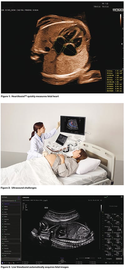
This study compares the performance of AI with that of expert clinicians who routinely assess fetal heart anatomy between 19 and 23 weeks of gestation. Specifically, the study focused on key views such as the four-chamber view, left and right outflow tracts, and the three-vessel view. This research involved 120 consecutive low-risk singleton pregnancies undergoing second-trimester ultrasounds. The findings indicate that technologies like HeartAssist™ can achieve accuracy comparable to expert visual assessments, suggesting their potential utility in evaluating fetal heart structures during the second trimester. In simple terms, the study showed that AI system and clinical experts have high levels of agreement when it comes to identifying the four fetal cardiac views in the study. This means that AI is nearly as effective as clinical experts in evaluating these views. Such findings highlight the potential of AI to support healthcare professionals by providing reliable diagnostic assistance, although it is important to note that AI should complement rather than replace human expertise. This growing synergy between AI and clinical experts could enhance diagnostic accuracy and efficiency in medical practice. We leveraged the insights gained from this study to explore the integration of AI technology, HeartAssist™, into our busy ultrasound unit, which conducts over 29,000 ultrasounds annually, including approximately 4,500 anatomical scans and 400 fetal cardiac ultrasounds each year (Figure 1).
We were particularly intrigued by the potential of this technology to serve multiple purposes during our evaluation process. In a busy academic practice, it is essential to balance work efficiently while maximizing educational opportunities for team members, all without compromising patient care. This can be achieved by integrating education into daily clinical routines and leveraging technology. Creating a positive learning environment is crucial; this involves fostering clear communication, providing a balance of autonomy and supervision, and maintaining enthusiasm for teaching. By embedding education within the clinical workflow, academic practices can ensure that learning is continuous and aligned with patient care priorities. This approach not only enhances the skills of healthcare providers but also improves the overall quality of care delivered to patients. Incorporating AI technology into clinical settings has proven to be immensely valuable, as it enhances skill development for beginners and boosts productivity for experienced team members. Initially, there was some skepticism about the integration of this technology.
However, it quickly became apparent that AI serves not only as an impressive tool but also as an effective teaching aid and a significant time saver. By facilitating skill sharpening for novices and streamlining workflows for seasoned professionals, AI technology supports a more efficient and educationally enriching environment in clinical practices. By integrating AI technology into our workflow, we successfully demonstrated its dual role as both a teaching tool and a catalyst for embracing technological advancements among our team members. Considering the concept of uncovering opportunities through challenges, we initiated discussions to introduce AI technology, which we believe would be highly beneficial (Figure 2). This integration allowed sonographers to receive real-time educational support while performing their tasks, thereby enhancing their skills and efficiency. Importantly, we were able to illustrate to the team the tangible benefits of adopting this technology, which included a significant reduction in the time required to evaluate, identify, and label fetal cardiac images, averaging a 5 to 10% decrease per patient.
This improvement not only streamlined operations, but also highlighted the value of AI in enhancing both educational and practical aspects of our practice. Some additional aspects of implementing this technology also were brought to light. When we inquired from our team about the utilization of the technology, we were pleasantly surprised to hear about additional benefits that we had not considered prior to implementation. It allowed the users to create a self base method with the technology to build confidence as well as skill level. The more seasoned sonographers also voice that they felt that the system was a fun way to test their skills against the artificial technology.
Future State
Our findings, along with those from other centers, demonstrate that the AI technology not only has a synergistic effect but also an additive effect. This means that when combined with existing methods, the technology enhances overall outcomes by working in harmony with current practices and contributing additional benefits. This dual impact underscores the potential of integrating advanced technology into various fields to improve efficiency and effectiveness of humans. We are continuously evaluating AI software to enhance both our workflow and educational goals across various levels of expertise. AI has the potential to significantly improve personalized learning by tailoring educational experiences to individual student needs, accommodating different learning paces and preferences.
In terms of workflow enhancements, AI streamlines various tasks, optimizes resource allocation within educational environments. At our facility, we adopt a step-by-step approach to integrating AI into our workflow. Initially, we assess the AI system in a dedicated workstation, separate from our routine operations. This “offline” evaluation method enables us to thoroughly test the AI software without disrupting our regular processes. By using typical prenatal images within the program, we can effectively assess its performance and capabilities in a controlled environment. We are currently assessing Samsung’s Live ViewAssist™ AI deep learning technology within this controlled environment. This advanced system automatically recognizes the specific views needed during live scanning and after performing a quality assessment of specific Using the Live ViewAssist technologies may reduce scanning keystrokes an average of 89%, streamlining processes, improving workflow efficiency, and reducing user variability in fetal imaging views, will extract them without any user interaction (Figure 3).
For the views extracted, the system will automatically annotate 47 different anatomical structures with up to 92% accuracy7. Live ViewAssist will also measure 46 different anatomical structures using the licensed ViewAssist™ | BiometryAssist™ | HeartAssist™ technologies7. This setup allows us to thoroughly evaluate the technology’s capabilities and effectiveness in a controlled setting. We have been pleased with the findings so far. These features have the potential to reduce scanning time and enhance consistency by minimizing user variability. Additionally, features such as EzCheck™ monitor exam progress, while EzReport™ provides instant access to measurement results. Using the Live ViewAssist technologies may reduce scanning keystrokes an average of 89%7, streamlining processes, improving workflow efficiency, and reducing user variability in fetal imaging. We will be rolling this program out to our team in a metered fashion to see if the same potential will translate to the ultrasound exam in our busy clinical environment.
Conclusion
The integration of Live ViewAssist AI deep learning technology into our workflow demonstrates significant clinical and operational benefits. By automatically recognizing and extracting specific views during live scanning, the system reduces scanning time and enhances consistency, minimizing user variability. This technology supports tools like BiometryAssist, ViewAssist, and HeartAssist, and features such as EzCheck and EzReport further streamline processes by monitoring exam progress and providing instant measurement results. As we continue to evaluate its effectiveness in a controlled environment, we are optimistic about its potential to improve workflow efficiency and reduce keystrokes. We plan to gradually implement this technology across our team to enhance both educational and practical aspects of our practice, ultimately aiming to improve patient care in our busy clinical setting. The future of prenatal ultrasound with AI integration is incredibly promising, and we are excited to contribute to this transformative process.