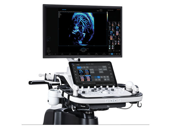
The HERA W10 Elite features Samsung's FreeForm™ design philosophy, engineered to minimize physical strain and create a natural workflow that adapts to your scanning needs.
IDEA AWARD 2018 Winner
The HERA W10 Elite earned the prestigious IDEA AWARD 2018 for its innovative ergonomic design and aesthetics, reflecting Samsung's commitment to enhanced user experience.
.
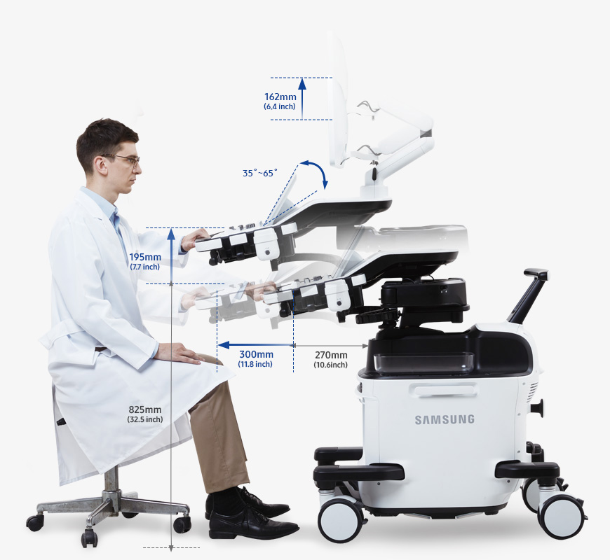
Control Panel Moving Mechanism
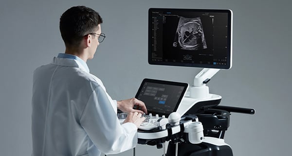
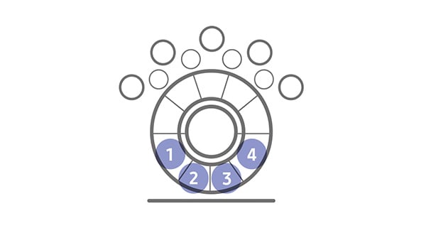
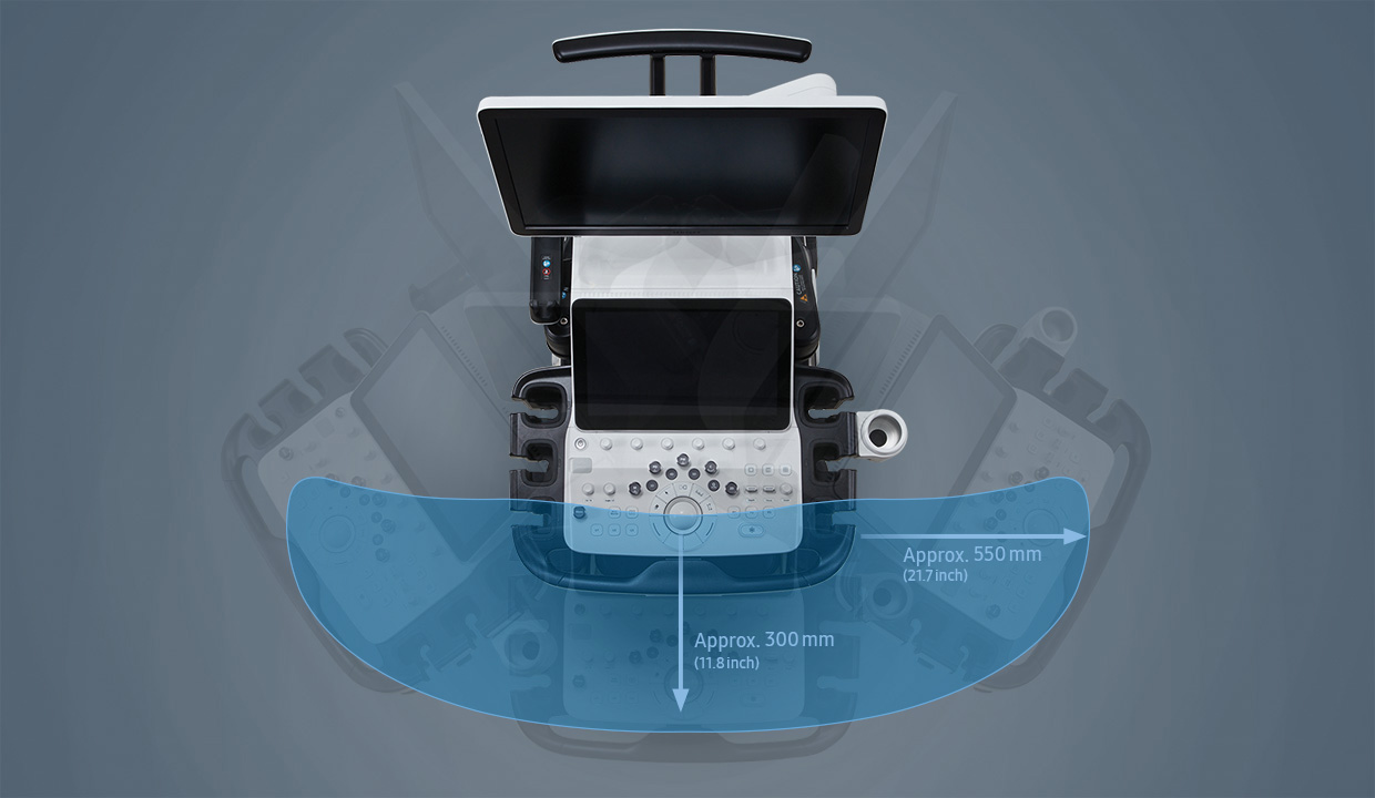
Samsung's Control Panel Moving Mechanism reduces shoulder stress by approximately one-third through improved positioning and simultaneous pull-and-rotate functionality, minimizing repetitive strain during extended scanning sessions.
* Control panel usability study compared to the Samsung WS80A. Tested using same body postures.